
7 Cardiology Lessons I Wish I’d Known as a New Grad
Valuble cardiology lessons for general practice

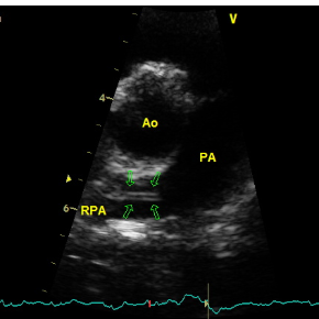
For many UK vets, heartworm disease remains something we read about rather than diagnose. However, increasing numbers of imported rescue dogs, international pet travel and changing climate patterns mean that Dirofilaria immitis is no longer exclusively a concern for colleagues overseas.
While the UK is not currently considered endemic for heartworm, imported cases are being encountered more frequently, and recognising the disease early can have a significant impact on outcome. Unfortunately, many affected dogs can appear remarkably well despite harbouring a potentially life-threatening parasitic infection. If you’re suspicious of heartworm in your patient and need help, we are here for you!
Any dog with a history of travel to, or importation from, endemic regions should raise suspicion. This includes many popular holiday destinations across southern Europe, including Spain, Portugal, Italy, Greece and parts of France.
Heartworm should be considered in:
Clinical signs can range from absent to severe, depending on worm burden and disease duration. Common presentations include:
In severe cases, large numbers of worms may obstruct blood flow through the right heart and vena cava, resulting in caval syndrome – a life-threatening emergency characterised by haemoglobinuria, weakness, collapse and cardiovascular compromise.
Diagnosis is often more complicated than simply running an antigen test.
Current American Heartworm Society recommendations advise both antigen testing and microfilaria testing when screening dogs. The combination improves diagnostic sensitivity and helps identify occult infections.
Commercial antigen tests detect proteins released primarily by adult female heartworms and are highly specific. However, false-negative results can occur in several situations:
The latter is particularly relevant in dogs that have previously received macrocyclic lactone preventatives. In these patients, circulating antigen may be masked and escape detection. Where clinical suspicion remains high despite a negative antigen test, further investigation is warranted.
The Modified Knott’s test remains a valuable adjunctive test.
A positive microfilaria test can support the diagnosis, identify circulating microfilariae and help detect cases that may be missed on antigen testing alone. Current Americal Heartworm Society (AHS) guidance recommends antigen and microfilaria testing together rather than relying on a single test modality.
Once heartworm infection has been confirmed, the next step is determining what damage has already occurred.
Thoracic radiography may reveal:
Echocardiography can be particularly useful for assessing:
Remember that it is often the secondary cardiopulmonary consequences of heartworm disease, rather than the worms themselves, that dictate prognosis and treatment requirements.
Treatment: the current gold standard
The American Heartworm Society currently recommends a multimodal treatment protocol consisting of:
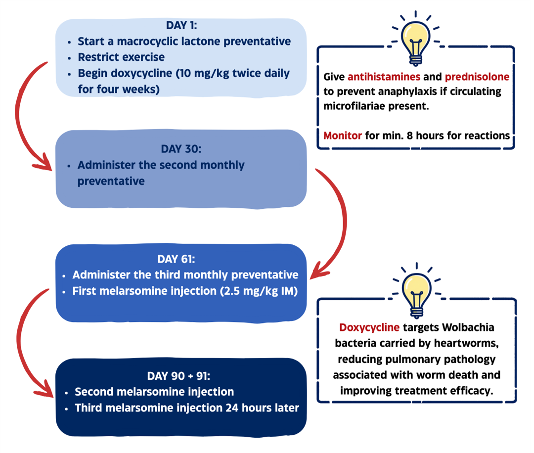
Prednisolone is often incorporated into treatment protocols to help manage pulmonary inflammation associated with dying worms, particularly in patients with more advanced disease.
One of the most important aspects of treatment is also one of the most overlooked.
As adult worms die, fragments can embolise into the pulmonary vasculature. Exercise increases pulmonary blood flow and significantly increases the risk of thromboembolic complications.
Strict exercise restriction should therefore be maintained throughout treatment and for at least 6-8 weeks after the final melarsomine injection.
Patients with caval syndrome or exceptionally heavy worm burdens may require mechanical extraction of adult worms before medical therapy can be safely undertaken.
These cases should be considered emergencies and referral is strongly recommended.
Heartworm may still be uncommon in the UK, but it is no longer rare. Any imported dog or patient with a history of travel to endemic regions should prompt consideration of Dirofilaria immitis infection.
Combining antigen and microfilaria testing, assessing disease severity carefully, and following current American Heartworm Society treatment recommendations can significantly improve outcomes.
If you have a patient with suspected or confirmed heartworm disease and would like support with cardiac assessment, echocardiography or management of secondary cardiopulmonary complications, the HeartVets team would be happy to help.
Valuble cardiology lessons for general practice

Veterinary Technician Specialists in cardiology – the inside scoop