
7 Cardiology Lessons I Wish I’d Known as a New Grad
Valuble cardiology lessons for general practice
As is the theme for most things in vet school, subjects are taught broadly, yet with enough minutiae to make you feel like they’re designed to trip you up in an exam. Then multiply that subject (and its sometimes huge variations) across multiple species. Cardiology is no different!
You may leave university with equal knowledge of tetralogy of Fallot, the intricacies of baroreceptors in systemic hypertension and myxomatous mitral valve disease physiology, but it can be surprisingly difficult to distinguish what is actually going to make a difference on day one of your first real vet job.
“With time, comes experience.” Training and working in the referral cardiology world has certainly given us plenty of that! So, let’s cut to the chase and impart a few wisdom nuggets that will hopefully help those starting out – and perhaps even bring a little practical clarity to those who are not so new to general practice.
It is very easy to give too much importance to whether a murmur has gone up a grade, or to a new loud murmur in a patient with no clinical signs.
Think of the heart as a musical instrument. Murmurs are caused by turbulent blood flow, and where that flow goes and the structures it interacts with on the way hugely influences how it sounds through your stethoscope.
We’ve had patients you can hear vibrating from the other side of the consult room with a tiny jet of mitral regurgitation that happens to roll around the left atrial wall and create a huge vibration on the left side of the chest, despite an otherwise normal heart!
As heart disease progresses, the structures of the heart change too, which can alter what you hear. So where can you practically apply this?
An older, small-breed dog with a moderate (grade III/VI) murmur may well have myxomatous mitral valve disease, and the American College of Veterinary Internal Medicine (ACVIM) guidelines recommend echocardiography to stage patients appropriately, regardless of clinical signs. If that grade III becomes a grade IV or V, it doesn’t mean a B1 patient is now B2 – auscultation does not replace echocardiographic assessment.
Conversely, a large-breed dog with a deep chest and a quiet murmur shouldn’t necessarily wait until the murmur gets louder before being investigated. Preclinical dilated cardiomyopathy (DCM) catches out many vets: murmurs can be surprisingly soft despite significant cardiac enlargement.
Treat the echo findings and relevant clinical signs – not the auscultation findings alone.
Echocardiography is arguably the most informative cardiac investigation, but it isn’t always practical to start there. When you’re deciding what to do next, first ask yourself: what am I actually trying to find out, and will the result change my clinical plan?
You cannot diagnose a cardiac disease with a stethoscope, even when the patient ticks every box for the most likely condition. Even we get caught out by auscultation findings that sound familiar!
Auscultation is an incredibly useful screening tool, but it is still just that – a screening tool. Even basic echocardiography can make a huge difference to diagnosis and treatment decisions, and may even save an owner paying for medication their pet doesn’t actually need.
Many cardiac patients also have concurrent respiratory disease. Advanced cardiac disease can exacerbate existing respiratory problems – particularly in a little old dog with a big left atrium and collapsing airways. Patients can also develop kennel cough, asthma or any number of other respiratory conditions.
Yes, frusemide may improve the cough, but that doesn’t necessarily mean the patient is in heart failure and it is the right treatment, or that there isn’t another underlying disease that needs addressing.
If a patient has a normal sleeping respiratory rate (SRR), or no change from their established baseline, active pulmonary oedema becomes much less likely. When the clinical picture doesn’t add up, investigate rather than automatically reaching for more diuretics.
We are constantly amazed by our feline patients! A surprising number of cats can have no clinical signs and completely unremarkable auscultation before presenting with some of the most severe echocardiographic changes we’ve seen.
Not all hypertrophic cardiomyopathy (HCM) patients have a murmur. ProBNP can be particularly useful here: screening relatives of cats with HCM or investigating before potentially destabilising events (such as anaesthesia or fluid therapy) can be invaluable.
For assessing the risk of left-sided congestive heart failure and monitoring pulmonary oedema, this simple measurement is a game changer. While gold-standard investigations such as thoracic radiography and echocardiographic assessment of filling pressures provide more definitive information, they aren’t practical to perform regularly.
Monitoring SRR trends gives owners a simple way to assess their pet at home and allows changes to be picked up much earlier. It is one of the easiest ways to have an extra pair of clinical eyes whilst empowering owners to engage with monitoring for disease progression.
We get it. It can be scary to ask for help, particularly when you feel like you “should” know the answer, or you’re not completely confident in your final diagnosis or treatment plan.
But there is absolutely no such thing as a stupid question! One of the things we are proudest of at HeartVets is being approachable and accessible. We love teaching and empowering vets and nurses in practice, whether you want to run a case past us for a sense-check or get guidance right from the start. Head to our telemedicine page for all things case advice, ECG diagnosing, radiograph interpretation and echo image checking at https://heartvets.co.uk/telemedicine/
So if your next cardiac case leaves you thinking, “I’m not quite sure about this one…” – ask. You might be surprised how much you learn.
And that’s probably the biggest lesson we’d give any new grad: you don’t need to know everything. You just need to know when to ask.
Valuble cardiology lessons for general practice
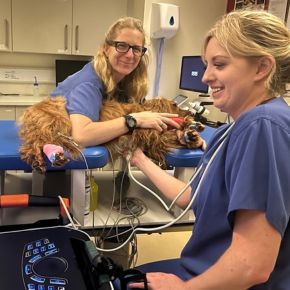
Veterinary Technician Specialists in cardiology – the inside scoop