Pacemakers are lifesavers for many of our canine patients, and whilst the knee-jerk reaction is often to avoid future anaesthetic procedures altogether, you may be surprised to hear that it is more straightforward than you think. Below is a concise, step-by-step framework you can follow the next time a pacemaker patient lands on your surgical list.
Contact us for telemedicine or referral support
1. The pre-op checklist
a. Clarify the procedure
- Is MRI involved? Many modern devices are MRI-adaptive, but only if specific settings are adjusted. Drop us a quick line before booking the scan so we can confirm compatibility and advise if anything needs changing.
- Thermocautery should not be used in these patients, so you may need to plan ahead for standard haemostasis.
b. Check the timing
Has the pacemaker been fitted in the last 10 weeks? Unless it’s an emergency, postpone elective anaesthesia. During this “bedding-in” window the lead tip is still fibrosing into the right-ventricular endocardium and infection risk remains higher.
c. Review the last interrogation
If a routine cardiology check-up is due, schedule it before the anaesthetic. We’ll confirm pacemaker battery status, capture thresholds and importantly – screen for any evolving structural disease that might require you to tweak your drug choices.
d. Rule out (or identify) structural heart disease
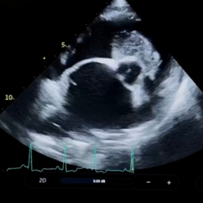
Fitting a pacemaker does not mean that patients cannot develop other structural cardiac disease such as degenerative mitral valve disease (DMVD) or other rhythm disturbances (such as atrial fibrillation). A focused echo to assess left-atrial size and systolic function will flag patients who may need an adjusted approach.
2. Choosing your drugs
Scenario | Premedication | Induction |
---|---|---|
Unsure of cardiac status/emergency | Opioid of your choice | Propofol or alfaxalone to effect, plus midazolam |
Minimal or no structural disease | Acepromazine IV and opioid | Propofol or alfaxalone to effect |
Documented structural disease | Opioid of your choice | Propofol or alfaxalone to effect |
Note: Alpha2 agonists (e.g. medetomidine) are best avoided; their vasoconstriction cannot be balanced by a reflex tachycardia.
3. Intra-op tips
- Jugular sampling – the right vein was ligated during pacemaker implantation, so opt for the left wherever possible.
- Once anaesthetised and the patient is still, the rate-adaptive pacemaker will default to its lower rate limit (60 bpm). Heart rate will not correlate with anaesthetic depth or nociception, so continuous bloods pressure, respiratory rate assessment and capnography should be used instead.
- Movement of the patient such as intubation, repositioning or CT gantry shifts triggers the rate-response sensor which will increase the heart rate. Don’t be alarmed; it settles when the patient is still again.
- Fluids – in patients with left atrial enlargement or poor systolic function keep to maintenance rates (or less) unless there is hypotension. But even then, be careful of boluses – the pacemaker will not mimic a physiological change in heart rate to compensate, and overperfusion is a real risk.
- Crash drugs – adrenaline remains your first-line for causing vasoconstriction and centralising the circulating volume. Note that heart rate will remain unchanged, which in some ways can be beneficial. KEEP AN EYE ON PULSE (make sure pacemaker is generating a pulse) if respiratory arrest occurs – even though heart rate may be stable, it may not be effectively oxygenating myocardium.
4. Post-op considerations
- Consider an early pacemaker check if the procedure inadvertently involved electrocautery, MRI, or any vigorous manipulation near the pacing lead.
- Antibiotics: your standard protocol will suffice, but if the original implant was within three months, consider extending coverage – we can discuss this with you if you need advice.
Key take-home messages
- Pause and plan – create a patient checklist, double check anything you aren’t sure of with the patient cardiologist.
- Structure dictates strategy – echo findings drive your fluid and premed choices more than the presence of the pacemaker itself.
- Monitor what matters – with heart rate unchanging, rely more on invasive or Doppler blood pressure, capnography and SpO2 to judge depth and perfusion.
- Avoid alpha2s and the right jugular, caution with fluids.
If you’ve got a procedure for a pacemaker patient coming up, we would be happy to discuss it with you to help optimise your patient care. These patients are usually very stable (with a bit of forethought) and less hassle than you’d think!
Discuss your case with our team