How pimobendan can change echocardiographic findings
Avoid misclassification of mitral valve disease in dogs receiving pimobendan

Given that most cardiac disease we see affects smaller, older dogs with degenerative mitral valve disease, it’s unsurprising that many also have concurrent respiratory disease – and in some, this may even be the primary problem. And what about cats?
A classic dilemma is the coughing patient with a heart murmur. Is it cardiac or respiratory? Historically, a thoracic radiograph was seen as a way to kill two birds with one stone: assess the cardiac silhouette, calculate a vertebral heart score (VHS) and evaluate the lungs all at once.
While evidence of left atrial enlargement and pulmonary oedema on X-ray remains highly valuable for diagnosing congestive heart failure (CHF), radiography leaves you guessing when results are equivocal or apparently ‘normal’.
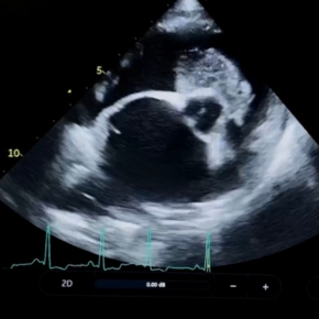
Obtaining high-quality thoracic images requires a still patient, inspiratory phase, and orthogonal views – ideally under anaesthesia with an inflated technique. But in a patient with a murmur, echocardiography should be the first step to classify structural disease, assess for pulmonary hypertension, and evaluate anaesthetic risk.
With appropriate stabilisation, many cardiac patients can safely undergo anaesthesia – but the question remains: are radiographs still your best option?
What may once have seemed the most accessible or cost-effective test can become a false economy if conscious radiographs fail to provide diagnostic clarity. A “normal” thoracic study does not guarantee a “normal” respiratory system.
So what are the other options?
If you’re unsure which test to choose or how to make advanced investigations work for your patients, we’re always happy to help.
Bronchoscopy allows direct visualisation of the airways from the trachea to the segmental bronchi. It’s particularly valuable in cases of chronic cough, airway collapse, tracheitis, bronchomalacia, foreign bodies, or dynamic obstruction.
Beyond inspection, bronchoscopy enables targeted sampling for cytology and culture, guiding specific therapy rather than repeated empirical trials. This can be the difference between months of steroids and antibiotics versus a swift, evidence-based resolution.
Advantages:
Bronchoalveolar lavage (BAL) is often performed alongside bronchoscopy, making it an efficient, single-anaesthetic diagnostic approach. It samples the lower airways to provide cytological and microbiological insight into otherwise invisible disease processes.
BAL is especially useful in suspected inflammatory, infectious, or allergic lung disease, helping differentiate eosinophilic bronchopneumopathy, bacterial bronchitis, parasitic disease, or neoplasia.
Why it matters:
Instilling fluid into the lungs of an anaesthetised patient can sound daunting at best, and it can be disheartening when pathology reports return as ‘non-diagnostic’. But with a little scope training and BAL practice, obtaining representative samples becomes a rewarding skill.
For those cases where referral is an option, our Wales patients can see RCVS Recognised Specialist Dave Dickson for cardiac assessment, bronchoscopy, and BAL – saving you all the stress of doing both!
CT has revolutionised respiratory diagnostics. By eliminating superimposition, it reveals fine detail invisible on radiographs and provides three-dimensional assessment of the thorax.
It is particularly useful for:
In patients with suspected neoplasia, inflammatory lung disease, or complex congenital anomalies, CT offers a definitive diagnosis and detailed disease mapping. Modern CT protocols enable rapid image acquisition under light sedation, minimising anaesthetic time and improving safety, even for compromised cardiac patients.
Advanced diagnostics aren’t just about precision – they’re about protecting patients from repeated empirical treatments, unnecessary side effects and ongoing disease. A clear diagnosis supports targeted therapy, faster recovery, and better long-term quality of life.
With improved anaesthetic safety and referral support, bronchoscopy, BAL, and CT are now more accessible than many clinicians realise – and often more cost-effective in the long run than a ‘quick set’ of radiographs.
The HeartVets cardiologists work alongside internal medicine teams in multidisciplinary hospitals, which is extremely beneficial for patients where input from both teams is required.
Take a look at our referral page to find your nearest referral service.

Avoid misclassification of mitral valve disease in dogs receiving pimobendan

Understand how left atrial size can cause a cough in dogs with mitral valve disease in our article.