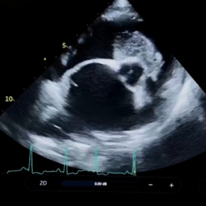
This article focuses on the presentation of acute left-sided congestive heart failure (CHF) in dogs and cats. Right-sided failure can also present acutely, but patients are generally more stable than those with pulmonary oedema and breathlessness. We are on the end of the phone if you are struggling to stabilise an acute case, or happy to see referrals for further investigations once stabilised.
It’s tempting to assume CHF in a dog with a known murmur and a pronounced cough, but concurrent or unrelated disease is common – and lung crackles alone aren’t diagnostic. Equally not all tachypnoeic cats have cardiac disease.
The key signs include:
- Elevated sleeping respiratory rate (SRR) – Ideally, owners of patients with known heart disease are trained to monitor SRR, so changes in trend are noticed early.
- Tachypnoea and altered respiratory pattern – Active pulmonary oedema causes tachypnoea. Rates >40 brpm in the clinic – especially if shallow, fast, or with paradoxical effort should raise concern. Orthopnoea (difficulty breathing when lying down, restlessness, or altered sleeping position) are often reported.
- A rapidly progressive cough – Coughing alone is not a sign of failure, but breathless dogs with pulmonary oedema may have a worsening cough.
- Left atrial (LA) enlargement – Significant LA dilation is usually expected with pulmonary oedema. TFAST provides a quick, low-stress assessment.
- Increased filling pressures – Doppler assessment of mitral inflow and related measurements (IVRT, pulmonary vein flow) help confirm CHF, but require confident and accurate echocardiography.
- Pulmonary oedema on thoracic radiographs – The best way to differentiate pulmonary oedema from other causes of dyspnoea, but should never be at the expense of patient stability. In stable patients, always obtain orthogonal views – start with DV before lateral positioning to avoid confusing atelectasis lung pattern with pulmonary oedema.
What can cause acute destabilisation?
Understanding the trigger helps direct therapy, improve outcome and manage owner expectations.
Common causes include:
- Progression of existing structural disease: Sudden events such as ruptured chordae or myocardial infarction may lead to rapid volume overload and require aggressive therapy.
- Change in heart rate or rhythm:
- Arrhythmias like atrial fibrillation (AF), supraventricular tachycardia (SVT) or ventricular tachycardia (VT) can destabilise previously stable hearts. Always get an ECG – some tachyarrhythmias aren’t obviously irregular.
- These rhythms can occur due to existing structural cardiac disease or can develop with minimal or no obvious structural disease, but cause tachycardia induced myocardial failure (TIMF) if sustained.
- Endocrine disease (e.g. hyperthyroidism in cats, hypothyroidism in dogs) can significantly affect cardiac function, and exacerbate structural disease, pushing patients into CHF. Screen for these where possible.
- Significant bradyarrhythmias (e.g., complete heart block, sinus node dysfunction) can eventually lead to CHF.
- NOTE: Electrolyte imbalances reduce success of arrhythmia control – check biochemistry when possible.
- Arrhythmias like atrial fibrillation (AF), supraventricular tachycardia (SVT) or ventricular tachycardia (VT) can destabilise previously stable hearts. Always get an ECG – some tachyarrhythmias aren’t obviously irregular.
- Environmental stressors – Stress and sudden life changes (especially in cats) can trigger decompensation and affect long-term stability, causing relapse if returning to the same environment.
- New medications or treatments – Steroids (especially injectable depot forms) commonly tip preclinical cats into CHF.
Recent fluid therapy or procedures involving IV fluids or drugs causing marked haemodynamic effects can also precipitate decompensation in dogs and cats.
Treatment approach
Your initial aim here is to treat the congestion (by clearing pulmonary oedema) and support cardiac output. Be sure to bear in mind the type of disease process occurring – dogs with volume overload (such as degenerative mitral valve disease, dilated cardiomyopathy) are VERY different to cats with hypertrophic cardiomyopathy (which is a disease of predominantly diastolic dysfunction).
- Hospitalisation:
- Ideally admit patients for stabilisation in a hospital setting – they can be stabilised more quickly and monitored more closely in the crucial stages. It allows correction of sudden changes and enables investigations in a calmer, stepwise manner
- Stress control: using butorphanol and creating a calm environment makes a huge difference in improving respiratory efficiency
- Oxygen: flow-by or oxygen tents can be useful providing these do not cause undue stress or excessive heating. Diuresis is more important in left-sided failure as patients do not tend to be hypoxic.
- Diuretics:
- Frusemide (first line): intramuscular (IM) or intravenous (IV) is best. For the first dose, do not prioritise restraint of a breathless patient for intravenous access if it is causing significant stress – give IM initially, then place an IV catheter when they are more stable.
- Start with 2mg/kg and adjust according to patient response. Repeating this hourly in dogs until respiratory rate is decreasing, or until the patient has received a total of 8mg/kg over 4 hours.
- IMPORTANT: be more cautious in cats –their blood pressure can drop too quickly, reducing the intracardiac, worsening diastolic function and causing a physiologic increase in heart rate which can worsen outflow tract obstruction and increase myocardial workload. Aim to drop down to 1mg/kg for subsequent doses, or increase the dose interval.
- If oral frusemide resistance is suspected, parenteral frusemide is still effective. Once the patient is stable, consider transferring to an alternative oral diuretic (such as torasemide)
- Effusions:
- Use POCUS to check for pleural or pericardial effusion, and drain these where significant (see our article on pericardial effusions for more information)
- Pimobendan:
- Used in patients with reduced systolic function to help support clearing congestion by improving forward function
- Do not use in patients with suspected/evidence of outflow tract obstruction
- IV preparation is available (but not licenced in cats), although this is an expensive single-dose. Instead give this orally once the patient is less breathless.
- Rate control:
- Sinus tachycardia is a NORMAL physiological response to advanced structural disease and CHF. This does not need correcting. If you aren’t sure, record and ECG and we can always help interpret it for you.
- If you have evidence of a tachyarrhythmia, aim for initial stabilisation of the patient with diuretics (+/- positive inotrope) first to improve respiratory rate before considering specific anti-arrhythmic drugs.
Summary
Before diving in and reaching for the frusemide, take a step back and double check the following:
- Are you confident this is CHF – not respiratory disease or something else?
- What’s the likely trigger? Tailor treatment accordingly.
- Are there complicating factors (e.g., endocrine disease, arrhythmia)?
These patients can be tricky to manage, particularly if you’ve taken over a case part-way through. It is always useful to go back to basics if patients aren’t responding as you may expect. And if you aren’t sure, we would be happy to help with case advice or referral.