
Cardiology cheat sheet – quick wins for everyday practice

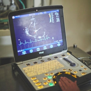
A team of veterinary cardiologists at work.

Short on time? These are the fast, high-yielding checks that make a real difference in a busy working day. Don’t worry if you’re still stuck, we can help with any case advice or interpretations of your investigations.

Find help here 


1) Auscultation Checklist

You can narrow most differentials and better focus your investigations if you consistently record the following. Noting these regularly also helps you and your colleagues detect meaningful change over time. For a succinct cardiac assessment, always document three key features:

  • Heart rate
  • Heart rhythm regular or irregular?
  • Murmurs and abnormal sound (gallop sounds, irregularities, absence of sound, muffling or dullness).

Describe: Which side? (left / right)

  • Which region?
  • How loud? (grade / intensity, can you feel a thrill?)
  • What timing? (systolic / diastolic / continuous)

Assess pulses during auscultation and describe:

  • Strength (strong / weak / bounding)
  • Timing with the heartbeat (synchronous / deficits)

When assessing the lungs, always record:

  • Breathing rate
  • Breathing effort (inspiratory / expiratory)

Lung sounds – describe:

  • Which side? (left / right)
  • Which region? (cranial / middle / caudal / dorsal / ventral)
  • What timing? (inspiratory / expiratory)

2) Radiograph Red Flags (in 60 Seconds)

Before interpreting patterns, give yourself the best possible chance by optimising technique.

  • Timing of exposure
  • Conscious or sedated (not intubated):  Aim for inspiratory timing. If this is not achieved, repeat the radiograph if possible – interpreting expiratory views significantly increases the risk of misdiagnosis.
  • Anaesthetised (intubated):  Aim for inspiratory timing with inflated views where possible.

Orthogonal views

  • There are very few justifiable reasons not to take orthogonal views in a stable patient.
  • Start with dorsoventral (DV) or ventrodorsal (VD) views before laterals to minimise atelectasis.

Correct orientation

    • Dorsoventral or ventrodorsal views: Cranial at the top, caudal at the bottom. Right side appears on the left of the image, left on the right.
    • Lateral views:  Cranial to the left, caudal to the right.

Key Features to Assess for Cardiac Disease

Cardiac silhouette – size and shape

    • Dogs: Large, rounded hearts raise concern for dilated cardiomyopathy (DCM), advanced myxomatous mitral valve disease (MMVD) or pericardial effusion. Be cautious in athletic working breeds, where this appearance may be ‘normal’.
    • Increased sternal contact or cranial bulging on a right lateral view may indicate right-sided enlargement. Remember the right heart is normally more prominent on a left lateral.
    • Cats:  Cardiomegaly tends to appear more “tall” than rounded, and the caudal vena cava may be obscured.
    • Consider calculating a vertebral heart score, bearing in mind breed variation.

Left atrium

    • A “hump” just caudal to the carina suggests left atrial enlargement.
    • In the absence of left atrial enlargement, primary left-sided congestive heart failure is very unlikely.
    • Marked left atrial enlargement may cause dorsal tracheal elevation.

Pulmonary vessels

    • Veins wider than arteries suggests venous distension in left-sided congestive heart failure (LCHF).
    • Arteries wider than veins may indicate pulmonary hypertension (PHT).

Lung pattern

    • Perihilar or caudodorsal alveolar pattern is typically associated with cardiogenic pulmonary oedema.
    • Patchy cranioventral or focal changes are much less likely to be cardiac in origin.

Pleural space

    • Dogs:  Pleural effusion suggests right-sided cardiac disease or a non-cardiac cause.
    • Cats:  Pleural effusion should place cardiac disease high on the differential list.
    • Need a second opinion? Submit your xrays here.

3) Speedy Electrocardiogram (ECG) Decoding

  • For the key elements, we don’t expect you to be calculating the electrical axis or measuring every wave. Cut to the chase with this general guidance: Ideally, record at least three leads. Always note paper speed and amplitude. Before attempting rhythm diagnosis, answer these fundamentals:

1. What is the overall rate?

    • Do not rely solely on the numbers given by your ECG equipment. Calculate manually where possible.
    • Decide whether the rate is appropriate for the patient’s environment and temperament.

2. Are the complexes regular or irregular?

    • Ignore morphology initially and assess the R–R interval alone.
    • It can help to cover most of the trace, leaving only the tips of R waves visible to assess regularity.

3. Can you identify sinus beats?

    • Look for a normal P QRS T complex with a consistent PR interval.
    • Decide whether sinus rhythm is present predominantly, intermittently, infrequently, or not at all.
    • If sinus beats are present, mark them – they provide a valuable reference.
    • If no sinus beats are identifiable, the arrhythmia is likely sustained.
  • Only once these steps are complete should you assess the arrhythmia in detail (and remember, more than one arrhythmia may be present).

P wave assessment (check all leads):

    • Are P waves visible?
    • If yes, are they associated with a QRS? Is the PR interval normal, shortened or prolonged? If P waves are not followed by a QRS, atrioventricular block should be suspected.

Assess abnormal QRS complexes:

    • Narrow complexes (similar to sinus beats) are more likely supraventricular or atrial.
    • Wide complexes (wider than sinus beats) are more likely ventricular in origin.
    • Note whether they occur as singles, couplets, triplets, runs or are sustained. If runs are present, calculate the rate for classification.

ECG Red Flags Requiring Prompt Action

  • Sustained or long runs of wide-complex tachycardia over 200 beats per minute: strongly suspicious of ventricular tachycardia and requires urgent treatment.
  • If the rate is below 200 beats per minute, consider accelerated idioventricular rhythm (AIVR). This is typically not dangerous, but if the patient is unstable or hypotensive, it is haemodynamically significant and still requires urgent treatment.
  • Sustained or long runs of regular, narrow-complex tachycardia (often over 200 beats per minute): suggestive of supraventricular tachycardia. This may not be immediately life-threatening but is often haemodynamically significant and can rapidly lead to cardiac enlargement and dysfunction.
  • Marked bradycardia  (particularly under 40 beats per minute): potentially life-threatening in conditions such as third-degree atrioventricular block, severe sinus node dysfunction or atrial standstill.

Want to check your diagnosis or need help with what to do next?

Give us a shout!

Read More