
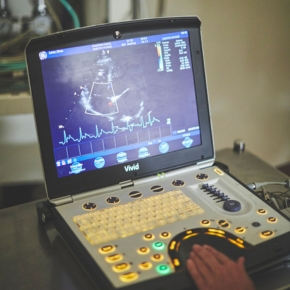
4 Reasons to Book an Echo Course this Autumn/Winter
From clinical confidence to career development, echocardiography is a skill that keeps on giving. Choose from our three upcoming courses to suit every level.
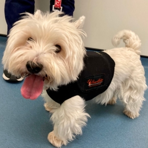
While hypertrophic cardiomyopathy (HCM) is a common consideration in feline cardiac disease, it rarely features on the radar in dogs. It is an unusual finding in our canine patients, but it can easily catch you out in practice, so read on to prepare for the unexpected. We are here to support you on your next steps when you’re ready.
The most recent study on HCM in dogs (2022) showed that affected patients are mostly small breeds, with a wide age range (though predominantly older dogs) and no sex predilection. This mirrors our experience in referral cardiology: while many breeds can be affected, we most often see it in terriers over 3 years old.
Most commonly, affected dogs are asymptomatic with a heart murmur. Some show signs of exercise intolerance, syncope, or hallmarks of congestive heart failure (CHF). If you are presented with an older, small breed dog with a heart murmur, the most tempting assumption is a diagnosis of degenerative mitral valve disease (DMVD).
Financial constraints or reluctance for diagnostics often result in dogs with suspected DMVD being started on pimobendan without further investigation. For dogs with HCM, dynamic outflow tract obstruction is common – precisely the type of case where pimobendan can be problematic.
Published date reports that 50% of dogs with HCM experienced sudden cardiac death under anaesthesia. Considering many older, small breed dogs require dental procedures in later life, this highlights the importance of cardiac investigations if a murmur is detected – even if the patient is clinically normal.
For those that undergo echocardiography, discovering an HCM phenotype can be alarming, and it may be unclear what to do next. Don’t worry — we’ve got you.
In dogs, HCM typically presents as left-sided, concentric, and symmetrical hypertrophy. If the heart appears thick on echocardiography, do not assume HCM immediately; a few other possibilities must be considered first.
Pseudohypertrophy: The heart may appear falsely thick if it is underfilled due to over-diuresis, severe dehydration, blood loss, or elevated right-sided pressures (pulmonary hypertension, congenital disease). Measure wall thickness in multiple locations and consider these factors.
In summary: review the entire echocardiogram carefully (or refer to a cardiologist if uncertain), obtain serial blood pressure readings (Doppler ideally), and run blood tests to exclude the most common secondary causes.
Data on outcomes is limited but aligns with our clinical experience. Sudden death under anaesthesia is the most common event. Other reported outcomes include syncope, arrhythmias, CHF, and sudden death outside anaesthesia. Unlike cats, thromboembolism is rare. Long-term survival is variable, ranging from 8 months to 14 years after diagnosis. Holter screening is particularly useful – you can rent one from our website here.
Do not reach for pimobendan immediately, even with left atrial enlargement. Case-specific discussion is essential. For dogs in CHF, diuresis is appropriate – but remember, HCM is not a volume-loading condition, so treat similarly to cats. Screening for arrhythmias with Holter monitoring is key, even in patients not yet showing syncope.
Remember, if something isn’t quite adding up or you want to discuss treatment options, we are here to help with case advice.
Learn about our telemedicine service
From clinical confidence to career development, echocardiography is a skill that keeps on giving. Choose from our three upcoming courses to suit every level.

We offer a variety of learning opportunities throughout the year – now’s your chance to let us know what, where and how you want to learn.