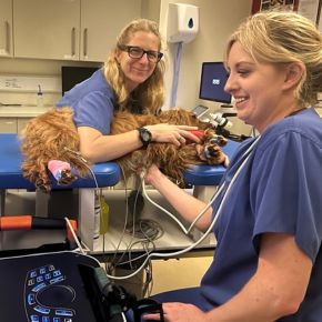
Prep and Go Checklist: Pericadiocentesis

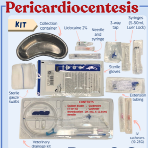
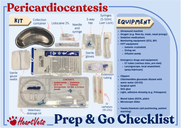
Part of a diagram showing a variety of items needed for a Pericardiocentesis kit

Our Prep and Go Checklists (emergency kit lists) are designed to help teams quickly gather everything they need, saving valuable time and reducing errors in critical moments. Veterinary nurses are perfectly placed to lead their use — coordinating care, anticipating needs, and keeping everything running smoothly. These resources are free to download, print and share with your whole team.
Download now


TOP TIPS: Pericardiocentesis

KIT (to use alongside our Prep and Go Checklist)   

  • Prepare a grab-and-go emergency kit in advance  
  • Ensure emergency drugs and equipment or crash box are available  
  • Set up oxygen supplementation as needed  
  • Prepare monitoring equipment (blood pressure, ECG, etc.)
  • Have warming aids ready

PATIENT

Before

  • Handle gently to minimise stress and cardiovascular compromise
  • Provide oxygen supplementation where tolerated and place an intravenous catheter
  • Clip and aseptically prepare the right thorax (typically 4th–6th intercostal spaces)
    • The right side is preferred for access and to reduce risk of coronary vessel damage
  • Consider intravenous fluid therapy (e.g. 5 ml/kg over 15–20 minutes) if hypotensive
    • Supports right heart filling, particularly in cardiac tamponade

During

  • Continuous ECG monitoring is essential:
    • Contact with the myocardium may trigger ventricular arrhythmias
    • Have lidocaine available for ventricular tachycardia
    • Be aware of electrical alternans (variation in QRS amplitude as the heart ‘swings’ within the effusion)
  • Monitor blood pressure closely
    • Hypotension is common due to impaired cardiac filling and should improve quickly after drainage
  • Collect and assess an initial fluid sample:
    • Check for clotting
    • Measure packed cell volume (PCV) and compare with peripheral blood (effusion PCV should be lower)

After

  • Continue close monitoring of heart rate and respiratory parameters – watch for recurrence
  • Maintain ECG monitoring during recovery to detect arrhythmias
  • Apply a small adhesive dressing; provide a spare for owners if needed

If effusion is not fully removed, residual fluid will drain into the pleural space and mild site leakage can occur from the thoracocentesis site – flag this to owners to manage expectations accordingly.

Download our Pericardiocentesis kit diagram


Read More