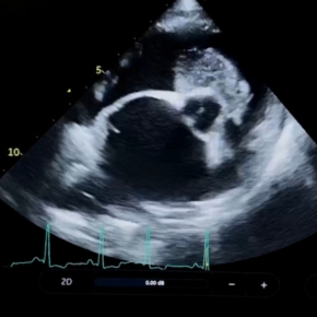
Even for the well-trained eye, early cardiac disease doesn’t always leave obvious clues. Subtle murmurs, fleeting arrhythmias, or even a perfectly normal exam can mask significant pathology.
The best veterinary care is proactive: spotting those early warning signs can extend – and sometimes save – lives. Here’s how to recognise the small clues in your clinical examination of asymptomatic patients that can make a big difference. Remember – if you aren’t sure, we are here to guide you if you need help.
DOGS
The soft murmur in an adult large-breed dog
When you’re not listening for a sign of mitral valve disease in a small breed, it’s easy to dismiss a low-grade murmur in a big dog with no clinical signs. But dilated cardiomyopathy (DCM) often starts silently – and the preclinical phase is where vets can make a huge impact.
In DCM, early left ventricular enlargement leads to stretching of the mitral valve annulus, creating a subtle systolic leak. The regurgitant jet is usually central, not causing vibration of the atrial wall. Particularly in a deep-chested dog, the sound may be faint or distant on auscultation, making it easy to miss.
Why early detection matters:
- Better outcomes: The PROTECT study (Summerfield et al, 2012) showed that treating preclinical DCM delays onset of signs and extends survival in Dobermanns.
- Preventing sudden death: In breeds like Dobermanns, ventricular arrhythmias can develop months to years before echo changes. Similarly, giant breeds such as Irish Wolfhounds can develop atrial fibrillation and ventricular arrhythmias. Holter monitoring detects this risk early, reducing the chance of collapse or fatal arrhythmia.
- Owner empowerment: Educating clients about DCM and its progression builds trust and prepares them for future decisions, both clinical and financial. There are some helpful owner information sheets that you can print out, which are available on our website.
- Holistic patient care: Knowing about DCM informs anaesthesia, fluid therapy, nutritional advice, and management of concurrent diseases.
- Responsible breeding: Feeding back early diagnoses to breeders and opting not to breed from affected dogs helps reduce incidence of this inherited disease.
What next:
- ProBNP blood test: Useful screening tool in some breeds (with published reference intervals). Significantly elevated proBNP should prompt echocardiography.
- Echocardiography: The only way to confirm the cause of a murmur and assess cardiac size, function, and wall thickness.
- ECG / Holter monitoring: ECG is useful, but a 24-hour Holter provides a more accurate representation of heart rate and rhythm away from the clinic to screen for intermittent arrhythmia. You can rent these from us – it’s easier than you think!
- Bloods and blood pressure: Identify concurrent issues like hypothyroidism or renal disease that could alter management.
- Diet review: Some specific diets have been linked to DCM, and patients may benefit from specific supplementation where there is proven insufficiency.
- Tailored treatment: Pimobendan is indicated where there is echocardiographic evidence of preclinical DCM. Specific anti-arrhythmic medication may be indicated, but the choice of drug will depend on echo findings and Holter results.
- Owner education: Following diagnosis of preclinical DCM, teach clients to monitor sleeping resting respiratory rate to obtain baseline values, and schedule echo or Holter rechecks every 6–12 months to look for progression.
Arrhythmia or abnormal heart rates
Just as ventricular arrhythmias in large-breed dogs with preclinical DCM can appear long before a murmur or echo changes, the same is true for ARVC in Boxers and English Bulldogs.
Atrial fibrillation (AF) can result from structural cardiac changes in small or large breeds – older or more sedentary patients may still appear ‘normal’. AF in large breeds can occur in isolation but can also precede the DCM phenotype in some giant breeds sch as Irish Wolfhounds, and increases the risk of sudden death. Single SVPCs or VPCs may signal underlying structural changes, from acquired disease to neoplasia. Even a rhythm that sounds regular (such as supraventricular tachycardia (SVT) or accelerated idioventricular rhythm (AIVR) may have a rate which is inappropriate for the patient situation. What sounds like sinus arrhythmia may be inappropriate for the patient (not young, fit or athletic breed) and may indicate a change in vagal tone or sinus node dysfunction. Don’t forget to feel for pulse presence and quality during auscultation to note any discrepancies.
What next:
- ECG: Essential to categorise the arrhythmia, but insufficient in isolation.
- Holter monitoring: More sensitive for intermittent arrhythmias and for assessing heart rates away from the clinical setting. Order yours here.
- Echocardiography: Almost always indicated after documenting an arrhythmia to evaluate cardiac structures and guide treatment.
- Blood tests: Helpful for identifying metabolic or systemic causes, depending on the arrhythmia classification.
CATS
Gallop sounds
Often mislabelled as a gallop ‘rhythm’, a gallop sound is the auscultation of a third heart sound caused by changes in myocardial compliance or intracardiac pressure. Even in the absence of a murmur, a gallop is rarely normal and should prompt further investigation.
Why it matters:
- Cats are masters of disguise – even those with advanced disease or imminent CHF can appear bright and well.
- Identifying preclinical disease allows treatment aimed at reducing thromboembolic risk in patients with echocardiographic evidence of increased left atrial size and reduced function.
- An apparent gallop sound my actually be an arrhythmia – further investigation can identify cats at risk of myocardial hypoxia and infarct (‘heart attack’).
- Knowing the cardiac status can inform safer anaesthetic and fluid therapy plans.
Normal auscultation findings (!)
Not all cats with cardiac disease have a murmur, arrhythmia, or gallop to give an early warning. While many preclinical cases may still be missed, there are two cohorts’ worth extra vigilance:
- At-risk cats: breeds such as Maine Coons, Sphynx, British Shorthair and Ragdolls, or cats related to those with known cardiac disease. Although any age can be affected, middle-aged and older cats are at greatest risk.
- Patients undergoing anaesthesia or IV fluid therapy (particularly middle aged and older cats): screening in these situations can prevent serious peri-anaesthetic complications.
What next:
- proBNP: A useful screening tool to flag underlying disease, although it does not equate to disease severity and false negatives mean it should not be used as a standalone diagnostic test.
- Resting ECG: To check for ventricular arrhythmia (which typically indicated myocardial disease in cats) and assess for ST segment changes.
- Echocardiography: The most accurate way to assess for structural disease and guide management.
- Bloods and blood pressure: Crucial for detecting concurrent conditions such as hyperthyroidism, renal disease, or systemic hypertension that can affect cardiac function or influence treatment.
Key Takeaways:
- Listen for subtle signs: quiet murmurs and arrhythmias can be indicators of serious underlying disease.
- Early detection is key for preclinical treatment, which can significantly improve patient outcomes. Referral to a cardiologist in the first instance may help to direct long term care by yourselves more efficiently.
- Owner education matters: informed clients are more engaged, better prepared, and more likely to follow proactive care plans.
- Follow up is important: cardiac disease can emerge, and changes can occur at any time. Patients can be stable for longer, with early warning of imminent deterioration with periodic echocardiography and Holter checks.