How pimobendan can change echocardiographic findings
Avoid misclassification of mitral valve disease in dogs receiving pimobendan

Rizzo’s owners noticed she was unwell when she was only 10 weeks old. She was breathing faster, with more effort and just wasn’t her usual bouncy self. Rizzo was seen at an out of hours emergency clinic who found she had a very loud heart murmur, her gums were pale, her lungs sounded crackly and she was struggling to breathe. The vet performed an ultrasound scan of her chest and heart and found her to be in congestive heart failure with fluid on her lungs (pulmonary oedema). Given Rizzo’s age the vet thought she was most likely to have a congenital heart defect that she would have been born with. Rizzo was stabilised overnight with oxygen, diuretic medication to remove the fluid from her lungs and a drug called pimobendan to improve the function of her heart. The following day she visited Mark Patteson at Vale Referrals for further investigations.
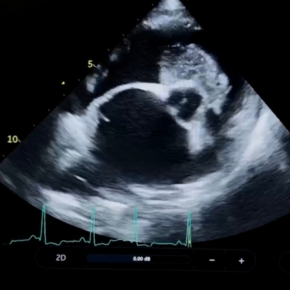
Using echocardiography (a heart ultrasound scan) Mark diagnosed Rizzo with a patent ductus arteriosus (PDA). The ductus arteriosus is small blood vessel that joins the aorta and pulmonary artery, which is needed before birth for blood to bypass the lungs and flow around the body providing oxygen from the placenta. At birth the PDA should close immediately as the puppy takes its first breaths. In some cases, the PDA fails to close. As there isn’t a valve in the PDA the blood can flow freely back and forth through it as the heart contracts and relaxes, causing a loud, continuous murmur. Puppies with a large PDA often develop an enlarged heart and congestive heart failure (fluid on the lungs/pulmonary oedema) in the first few months of life, and sadly most dogs left untreated will die before a year of age. This is why echocardiography of any puppy or dog with a loud murmur is important. We can assess the heart for the type, severity and prognosis all with one scan, usually without sedation.
Fortunately, we were able to close Rizzo’s PDA using a minimally invasive surgical procedure (“keyhole surgery”). Under general anaesthesia a small device called an Amplatz canine ductal occluder (ACDO) is passed via the femoral artery to the PDA, using fluoscopic guidance (like a moving X-ray image). Once released, the ACDO sits like a plug in the PDA, stopping blood flow and sealing the hole. Normal blood flow resumes in the great arteries and over time the heart size will reduce and the dog will be cured. Lucky Rizzo had this surgery performed in July this year, and all went smoothly.
We saw her a few weeks ago for her post operative recheck and her heart is getting back to its normal size and function. Rizzo’s owners say she has a new lease of life and plays non-stop!

Avoid misclassification of mitral valve disease in dogs receiving pimobendan

Understand how left atrial size can cause a cough in dogs with mitral valve disease in our article.