Holter monitoring in general practice
Elevate your cardiac case management with Holter monitoring in general practice

Holter monitoring in dogs and cats is often seen as a referral-only tool, but it’s more accessible than you might think. Forget the image of your old, bulky and somewhat temperamental ECG machine lurking at the back of a cupboard – modern Holters are lightweight, easy to use, extremely well tolerated and incredibly valuable in everyday practice. Here’s how this simple test can transform your cardiac case management.
Visit our website to learn more or to request a Holter rental today.
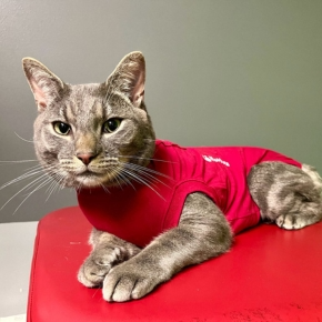
Despite the confusing name (often mistaken for “halter” – the headgear mostly seen in livestock and equine), the first Holter was invented by Norman Holter in the 1940s to record a human ECG whilst on the move. Today’s devices weigh as little as 35g and record 3 and 6-lead ECGs continuously for up to 14 days while the patient carries on as normal.
Conditions like dilated cardiomyopathy (DCM) and arrhythmogenic right ventricular cardiomyopathy (ARVC) carry a risk of sudden death, often due to complex ventricular arrhythmias – even in preclinical stages. These diseases typically involve two key elements: structural changes (seen on echo) and electrical disturbances (seen on ECG). Importantly, these don’t always appear together.
A patient might have a normal or mildly abnormal echo but still show serious arrhythmias on a Holter. A regular rhythm in the clinic doesn’t rule out problems at home – some of the most worrying ECGs come from patients with no structural changes and no clinical signs.
That’s why the gold standard for screening in DCM and ARVC is both echocardiography and 24-hour Holter monitoring. And it’s not just limited to these conditions – any myocardial disease, including myocarditis, advanced degenerative mitral valve disease (DMVD), or HCM, can cause ventricular arrhythmias.
Collapse can stem from cardiac, neurological, or systemic disease. Holter monitoring plays a vital role in ruling arrhythmia in or out as a cause – particularly before sedation or anaesthesia for further workup.
Capturing an event on Holter is ideal but not essential. Even without a collapse episode, longer recordings (48 hours or 7 days) provide insight into resting vs active heart rate, rhythm variability, and hallmark patterns like sick sinus syndrome (SSS). This can help differentiate between benign and dangerous rhythms.
Spotting an arrhythmia on an in-clinic ECG is helpful, but it doesn’t always tell the whole story. Treatment decisions often depend on the mean heart rate (in atrial fibrillation for example), which can be misleading in a stressed patient within a clinical setting.
Holter monitoring gives a more accurate picture of rhythm over time and helps assess clinical significance, particularly when arrhythmias may be secondary to non-cardiac disease (e.g. GI, respiratory or neurological). It’s a great tool for deciding whether and how to treat.
A patient may appear clinically improved on medication, but we can only confirm good control – and check for pro-arrhythmic effects by repeating a Holter after starting treatment.
Holter also supports long-term monitoring. In progressive disease, six or twelve-month checks (often alongside echo) can catch changes early. Some anti-arrhythmics may negatively impact systolic function, so Holter data helps guide medication adjustments as disease evolves.
Sometimes, echo findings raise flags – like a DCM phenotype in an unusual breed or left atrial enlargement with no obvious cause. These can be signs of tachycardia-induced cardiomyopathy (TICM), where sustained or frequent arrhythmias drive structural changes. Left untreated, this can lead to tachycardia-induced myocardial failure (TIMF).
Early arrhythmia control can stabilise or even reverse these changes. Similarly, conditions like tricuspid dysplasia are associated with atrial arrhythmias that may mimic structural progression or drive clinical signs such as weakness or collapse. Holter screening at diagnosis and points where there are structural changes is recommended.
Aortic stenosi is another example – often associated with exertional arrhythmias. These may not be evident in the clinic but can cause syncope or even sudden death if undetected. Holter monitoring provides crucial context in these cases.
Not at all! With Heart Vets, you can rent a Holter to use in practice – no special equipment needed. We’ll post everything to you (express shipping available), and fitting is simple enough to be done within the time of vet or nurse consult. Once returned, our wonderful analysts review the data, and our cardiology Specialists provide a tailored report with guidance for further testing or treatment. Our team is also on hand to support you if any issues arise.
Need More Info?
Visit our website to learn more or to request a Holter rental today.
Elevate your cardiac case management with Holter monitoring in general practice

Cardiac disease and endocrinopathies – what to do when things get complicated.