How pimobendan can change echocardiographic findings
Avoid misclassification of mitral valve disease in dogs receiving pimobendan

The following case study shows how Specialist referral helped clarify a complex presentation, allowing for a more tailored treatment plan that significantly improved his quality of life – just when euthanasia was being considered. With a fresh perspective and collaborative input, we were able to adjust medications, reduce side effects, and give the patient many more good days at home.
Rocket, an 11-year-old male neutered Jack Russell Terrier, was referred to Dave Dickson (one of our Directors and an RCVS Recognised Specialist in Cardiology), at HeartVets for optimisation of his cardiac treatment plan. He had a presumptive diagnosis of degenerative mitral valve disease (DMVD) and congestive heart failure (CHF) three months earlier, following a four-month history of non-productive coughing with terminal retch and reduced exercise tolerance. The cough had worsened in both frequency and severity, now occurring multiple times daily.
On initial presentation to the primary care vet, Rocket had a very loud (grade VI/VI) left-sided and moderate (grade III/VI) right-sided systolic murmur, with a thrill over the left apex and mild adventitious lung sounds. He was started on pimobendan and frusemide, which led to marked improvement in exercise tolerance and partial resolution of the cough. Polyuria and polydipsia were noted but manageable.
Several weeks later, Rocket collapsed while running – described as brief ataxia, followed by lateral recumbency and loss of consciousness, resolving spontaneously within 5 minutes. No arrhythmia or other abnormalities were noted on urgent veterinary examination. Though his cough persisted, it later worsened, becoming more frequent and prolonged.
His frusemide dose was increased, and sleeping respiratory rate monitoring at home showed rates mostly under 30 bpm, although occasional laboured breathing was noted. Conscious thoracic radiographs (limited due to inspiratory phase) suggested a possible interstitial/alveolar pattern. Despite further increases in frusemide and the addition of benazepril and spironolactone, Rocket’s clinical response was poor, and he appeared more subdued. Renal parameters worsened, coughing remained persistent, and he began having urinary accidents overnight. No further syncopal events occurred. With concern for declining quality of life and limited response to treatment, his owners agreed to referral as a final attempt to improve Rocket’s condition before euthanasia.
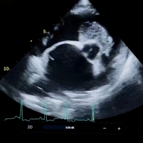
Dave performed conscious echocardiography, which confirmed degenerative mitral valve disease with an eccentric regurgitant jet tracking along the left atrial free wall. There was no evidence of left atrial or ventricular enlargement; the left heart appeared underfilled with normal estimated filling pressures from Doppler studies. In contrast, there was moderate right atrial enlargement, mild right ventricular dilation, septal flattening, and reduced pulmonary artery distensibility. Moderate tricuspid regurgitation and pulmonic regurgitations were noted without significant valvular pathology but significantly elevated pressure gradients. The remaining valves were normal, and there was no intracardiac shunt or evidence of pericardial, pleural, or abdominal effusion.
ECG showed showed sinus rhythm with no ectopic activity. Blood pressure was normal. Angiostrongylus SNAP test and faecal smear were negative.
The findings were suggestive of pulmonary hypertension (PHT), likely secondary to underlying respiratory disease, alongside early (mild) degenerative mitral valve disease. Based on echocardiographic findings, he met ACVIM stage B1 criteria; however, due to the use of frusemide and underfilled left heart, repeat imaging after a period without diuretics was advised for accurate staging.
With no evidence of cardiomegaly or CHF, frusemide was gradually reduced while monitoring sleeping respiratory rate (SRR), and sildenafil was initiated. Pimobendan was continued, pending reassessment (this is also a pulmonary vasodilator and provides right heart inotropic support). Rocket showed marked clinical improvement within a matter of days – he was brighter, more active, and playful. His cough persisted (but was milder and less frequent), and respiratory effort improved. Frusemide, benazepril, and spironolactone were discontinued as SRR remained stable (22–24 bpm).
Repeat echocardiography at four weeks showed a mild increase in left-sided dimensions reaching the upper end of normal (borderline ACVIM stage B1/B2), with no evidence of CHF. Right heart dimensions were largely unchanged, with a slight reduction in tricuspid regurgitation velocity. Renal parameters had improved on bloodwork.
Further investigations (inflated thoracic radiographs or CT, bronchoscopy, and bronchoalveolar lavage) were discussed to evaluate for primary respiratory disease. Due to Rocket’s clinical improvement and financial considerations, his owners declined further diagnostics unless signs recurred.
Rocket’s case highlights the challenges of managing patients with overlapping cardiac and respiratory signs – a scenario we see often at Heart Vets.
The referring vet’s initial concern was the very loud murmur, audible without a stethoscope and accompanied by a thrill. While this might suggest advanced heart disease, Rocket’s left-sided changes were mild. Eccentric mitral regurgitation jets can cause loud murmurs by vibrating nearby structures, especially in smaller or leaner dogs. It’s a reminder that murmur intensity doesn’t always reflect disease severity – and without echocardiography, we can’t determine the cause or clinical significance.
Heart rate and rhythm are also key clues. In suspected CHF, we typically expect sinus tachycardia and loss of sinus arrhythmia due to increased sympathetic tone. A murmur alone shouldn’t distract from these other vital signs.
Coughing is usually triggered by irritation of receptors in the larynx, trachea, or bronchi. In older small-breed dogs, common causes include chronic bronchitis, tracheal collapse, and bronchomalacia. Other differentials include infections (e.g. Angiostrongylus), secondary bacterial disease, or neoplasia. Mitral valve disease can contribute too – an enlarged left atrium may apply pressure dorsally the airways and further stimulate cough receptors. If there is evidence of significant left atrial enlargement on echocardiography (even in the absence of congestive heart failure), it is important to manage owner expectations as the cough is unlikely to change if there is no respiratory disease to address.
A ‘positive response’ to frusemide can mislead us toward a CHF diagnosis. Interestingly, whilst many of the mechanisms are still unknown, human studies show that frusemide has complex effects on the airway including prevention or attenuation of bronchospasm. This does not mean that frusemide is the correct choice for primary respiratory patients – there are more suitable drugs which do not have renal side effects which can be used more appropriately.
PHT is commonly secondary to left-sided congestive heart failure (CHF), but diagnosis and management can be complex – especially in patients with concurrent respiratory disease. It is essential to consider primary respiratory causes alongside cardiac investigations.
Sildenafil should not be used in CHF patients without first confirming that CHF is well-controlled, and the patient is clinically stable. A formal diagnosis of PHT (via Doppler echocardiography) is crucial, and owners should be informed of the potential risks of treatment.
In chronic CHF, pulmonary vasoconstriction can in fact help to protect the lungs. Using sildenafil (a pulmonary vasodilator) in unstable cases can cause ‘flooding’ of the lungs and acute destabilisation. Its use should be reserved for carefully selected patients, under appropriate guidance.
The collapse was likely to be linked to the pulmonary hypertension (left heart underfilling and reduced forward flow rather than arrhythmia, as no abnormal rhythms were detected on our ECGs. Holter monitoring would be useful to assess this further and rule out intermittent arrhythmia as a cause of the collapse and would be our next step should the collapse recur.
You can refresh your ECG knowledge with HeartVets at any time on our website.
Avoid misclassification of mitral valve disease in dogs receiving pimobendan

Understand how left atrial size can cause a cough in dogs with mitral valve disease in our article.